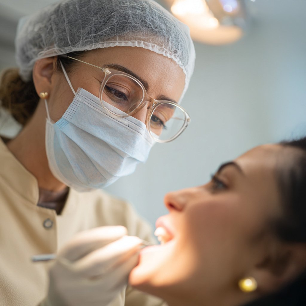
A surgical guide is a patient-specific 3D-printed stent for flapless implant placement in single- or multiple-tooth gaps. The guide is planned from digital records so that the osteotomy path and device position are set before surgery. Placement must follow the IFU for the selected implant and guide.
Planned osteotomy position: The patient-specific surgical guide is used to transfer the planned implant position into the jawbone.
Flapless protocol: When no flap is raised, sutures are not required. Soft-tissue disruption and post-operative swelling or pain are typically lower than with a flapped approach.
Immediate loading option: When the protocol allows, a prosthesis can be started without waiting for flap healing (immediate loading). Confirm primary stability and follow the IFU.
Digital implant planning steps
3D tomography: When CBCT slices support implant placement, an intraoral record is taken and both datasets are imported for planning.
3D implant planning: The records are reviewed in planning software to assess bone volume and quality. Prosthetic implant positions are set and checked on the 3D view before surgery.
Implant placement: The surgical guide is seated, the osteotomy is marked through the sleeve, the site is prepared, and the implant is placed on the planned axis. A healing abutment is then seated. Follow the IFU.
BROCHURES AND DOCUMENTS
SHORTER GUIDE
The surgical guide is used for flapless placement in single- or multiple-tooth gaps. It transfers the planned osteotomy to the site. Open the Shorter Guide brochure for the documented workflow.
CAD/CAM planning uses the digital library files for the selected implant connection. Download the digital library and matching scan-body files from the link.
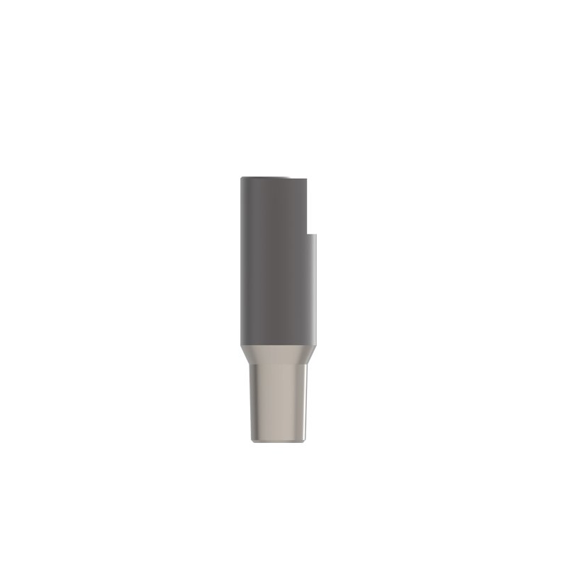
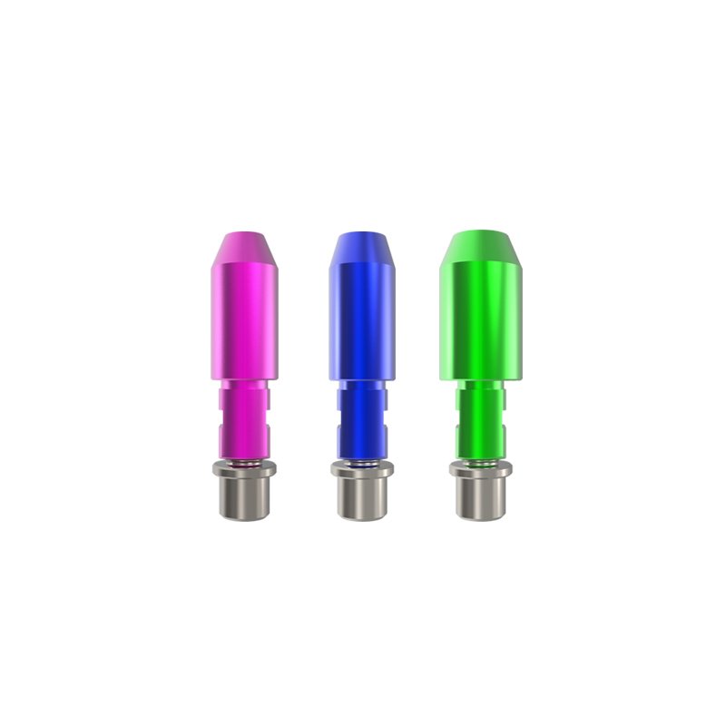
Scan bodies transfer the implant position into a 3D model for CAD/CAM. Use the scan body and digital library files specified for the selected Shorter connection. Restorative planning must follow the IFU for that component.
MULTI SCAN BODY
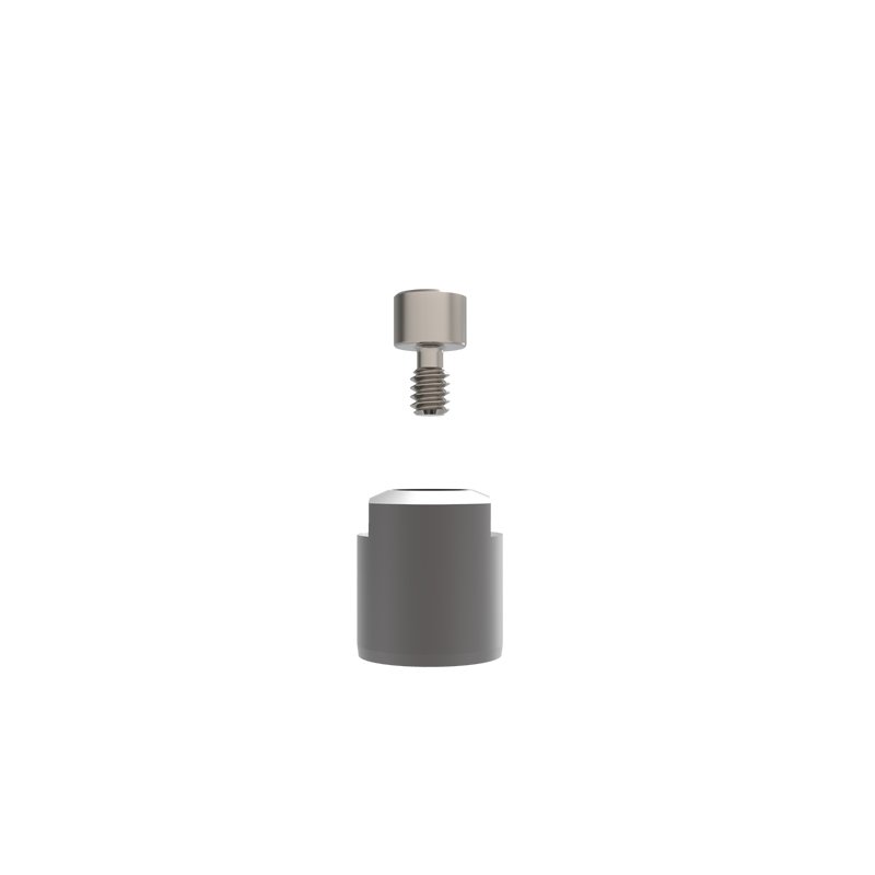
Scan bodies transfer the implant position into a 3D model for CAD/CAM. Use the scan body and digital library files specified for the selected Shorter connection. Restorative planning must follow the IFU for that component.
MULTI ANGLED SCAN BODY
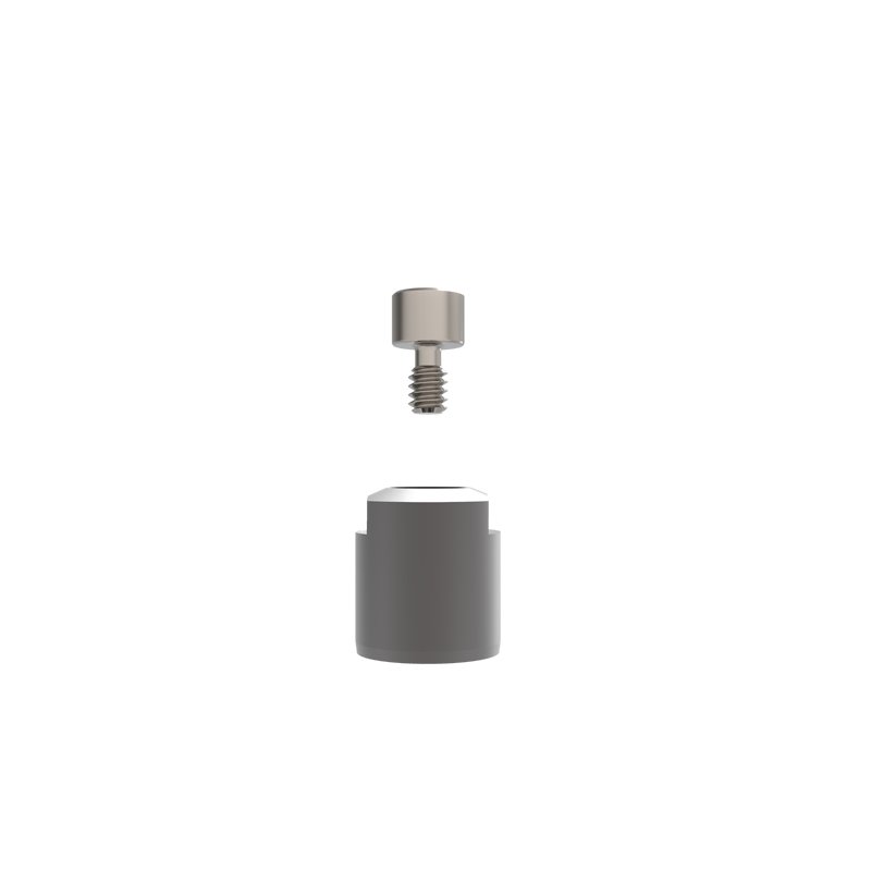
Scan bodies transfer the implant position into a 3D model for CAD/CAM. Use the scan body and digital library files specified for the selected Shorter connection. Restorative planning must follow the IFU for that component.
DIGITAL ANALOG
Scan bodies transfer the implant position into a 3D model for CAD/CAM. Use the scan body and digital library files specified for the selected Shorter connection. Restorative planning must follow the IFU for that component.
NTA S-PRO intraoral scanner
The NTA S-PRO intraoral scanner records color intraoral data and updates the scan as the wand moves:
Color intraoral scan: Hard and soft tissues are shown in color so they can be distinguished on screen.
Built-in anti-fog: The scan tip uses automatic heating to limit fogging so a pass can be completed without interrupting data capture.
Scan depth of field: Specified depth of field is 15 mm.
Cloud storage: Scan files can be stored in the cloud for transfer between clinic and lab.